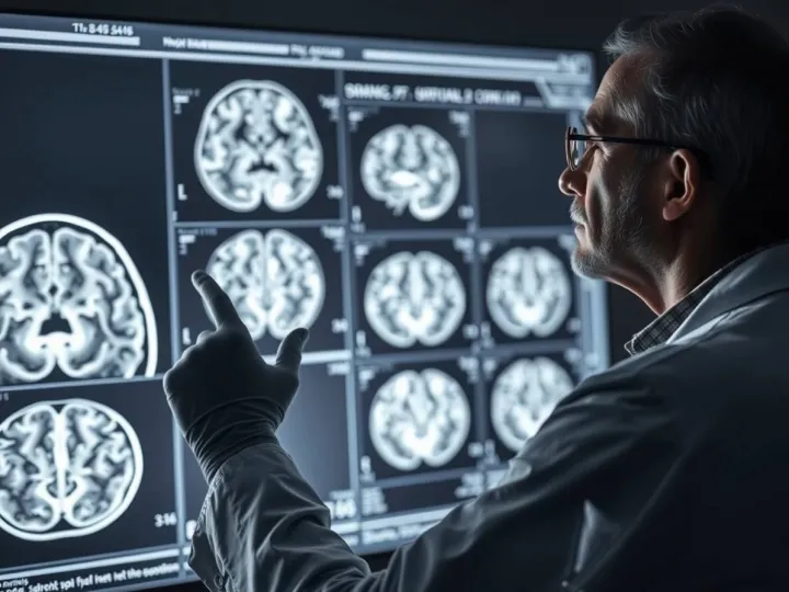
The study examined recordings from 29 individuals undergoing stereo-EEG for the assessment of drug-resistant epilepsy. While clinical focus traditionally centers on gray matter, where neuronal cell bodies generate the clearly recognizable activity of seizures, these recordings also capture signals from white matter tracts. White matter has historically been seen as a passive relay structure, supporting long-range connections without contributing substantially to measurable electrophysiological patterns. The availability of high-density intracranial recordings provided an opportunity to test this assumption directly by analyzing the electrical behavior of white matter during interictal periods, the transition into seizures, the seizure itself, and postictal recovery.
Across participants, white matter signal power showed a consistent signature. Although baseline activity remained lower than that of gray matter, white matter exhibited measurable changes across seizure states. Signal power decreased at the peak of the seizure but remained elevated following seizure termination. This pattern distinguishes white matter from cortical recordings, which tend to normalize quickly after ictal events. The dynamics suggest that white matter does not merely echo cortical signals but displays its own state-dependent behavior, particularly in the periods surrounding seizure propagation and recovery.
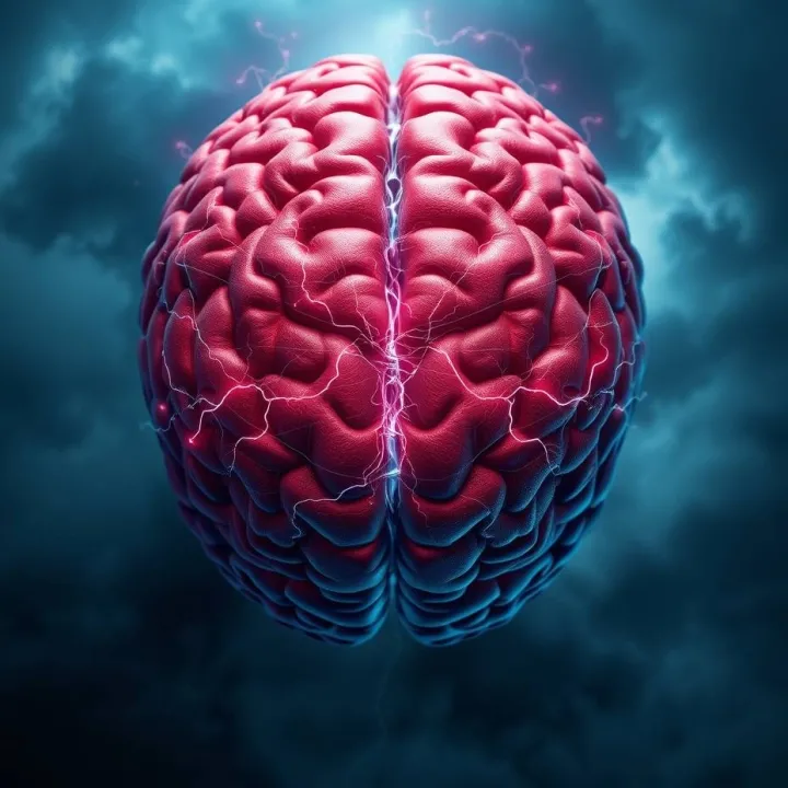
To understand how these signals relate to network communication, the researchers examined functional connectivity. During normal and preictal periods, white matter exhibited lower connectivity than gray matter, matching expectations about the relative richness of cortical processing. However, during seizures, this relationship reversed. White matter connectivity increased sharply, surpassing cortical values. These increases persisted after accounting for distance and spatial configuration of electrodes, indicating that they reflected genuine network relationships rather than simple volume conduction or proximity effects. The widespread rise in white matter connectivity suggests that seizure activity engages long-range communication pathways, with the white matter acting as a central conduit for information flow when the brain enters highly synchronized, pathological states.
A key part of the study involved integrating structural tractography with the stereo-EEG data. This allowed the authors to examine whether functional patterns observed in white matter corresponded to underlying anatomical pathways. Their analyses revealed that white matter functional connectivity during seizures aligns closely with the structural connections linking cortical regions. White matter electrodes showed increased coupling specifically along known fiber tracts, and the strength of these connections reflected the degree to which structurally connected cortical regions were communicating. Importantly, white matter connectivity did not simply correlate with other white matter tracts but largely mirrored cortico-cortical pathways. These findings reinforce the idea that white matter recordings reflect ongoing information transmission between cortical areas rather than independent white matter processes.
The study extended the analysis to clinical outcomes by comparing individuals who became seizure-free after surgery with those who continued to experience seizures. Patients with poor outcomes demonstrated significantly higher white matter connectivity during seizures, particularly in connections linking the presumed seizure onset zone to spatially distant pathways. In these individuals, seizure dynamics appeared less localized and more distributed across large-scale networks. This observation supports the distributed epileptic network hypothesis, which proposes that in some forms of epilepsy, seizures do not originate from a single discrete region but emerge from the interaction of multiple interconnected nodes. When a seizure is generated by a distributed network, removing one region surgically may be insufficient to disrupt the broader pathological circuit. The white matter thus provides a way to visualize how extensively the seizure onset zone communicates with remote brain regions, offering a potential marker for identifying patients who are less likely to benefit from focal surgery.
By capturing structural and functional properties simultaneously, the study offers a new vantage point for understanding seizure propagation. White matter recordings act as a window onto the pathways along which pathological synchronization travels. They also underline the importance of considering both gray and white matter in the clinical evaluation of epilepsy. Much of the current diagnostic framework relies on cortical measurements, yet the present results show that white matter may carry essential information about the spatial distribution and communication patterns of epileptic networks. These insights could support improved surgical planning, more accurate predictions of treatment outcomes, and potentially new strategies for neuromodulation that target communication pathways rather than isolated cortical regions.
Within the framework of Seven Reflections' Dimensional Systems Architecture, the findings illustrate how system behavior becomes visible not only at the nodes where activity is strongest but also in the connective fields that shape how information propagates. White matter functions as an architectural substrate that defines the system's communication patterns, and seizures represent a state in which the system temporarily shifts into a high-coherence mode across its connective layers. The fact that poor clinical outcomes correlate with elevated white matter connectivity echoes a core DSA principle: the integrity and behavior of a system depend not solely on activity within regions but on the structure and saturation of the pathways linking them. Understanding pathology therefore requires observing how information traverses the system's deeper architecture, not only where it emerges.
The study highlights a shift in how neuroscientists may conceptualize epilepsy, moving from a localized model toward a network-based understanding that incorporates both structure and function. White matter, once treated as a silent background element, emerges as a key part of the system's dynamic landscape, offering diagnostic and conceptual insight into how seizures unfold and why they resist certain interventions. As research continues, white matter electrophysiology may become a standard component of epilepsy evaluation, providing a more complete view of how the brain transitions into and out of pathological synchrony