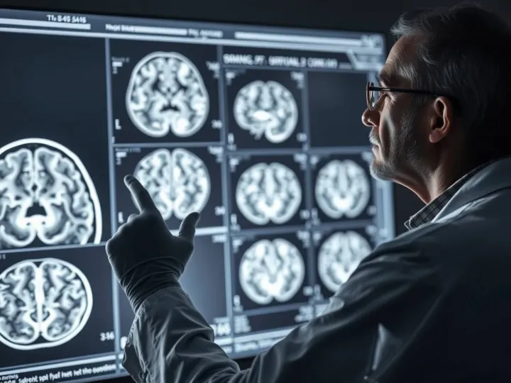
A large new study published in Cerebral Cortex (Open Access) provides an unusually detailed look at how brain wiring differs in autistic children and young adults. Instead of examining whole brain regions or broad networks, the researchers focused on the brain's "white matter" pathways - bundles of fibers that allow different regions to communicate. Using advanced mapping techniques, they were able to pinpoint subtle differences along very specific segments of these pathways.
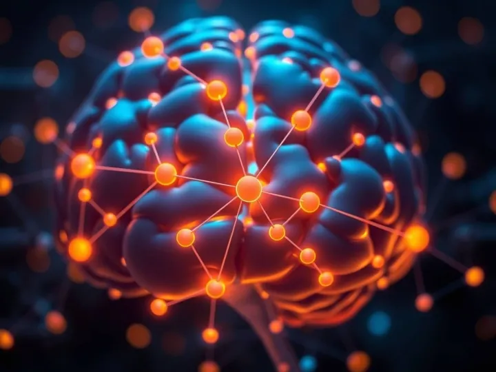
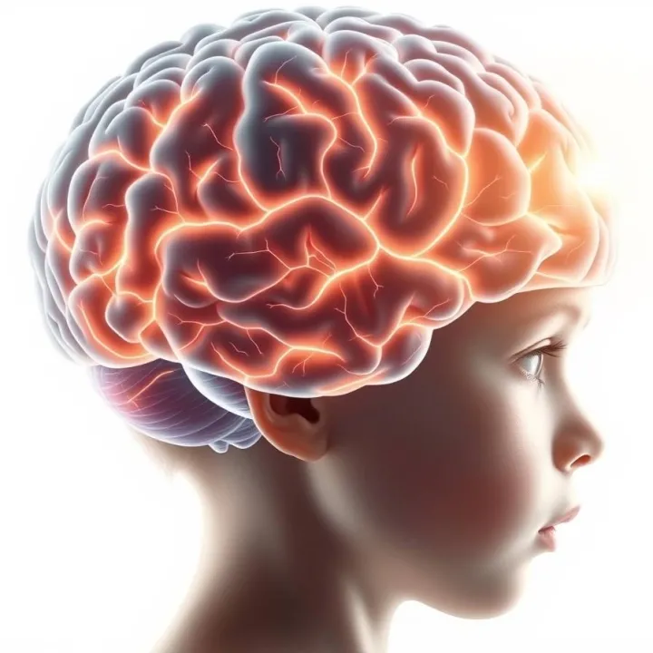
White matter tracts act like highways, linking parts of the brain involved in movement, sensation, language, emotion, and decision-making. Earlier research using diffusion MRI has found that autism is associated with differences in these pathways, but studies often disagreed about where these differences appeared. Traditional imaging tools average signals across large brain regions, which can blur small but important variations.
To overcome these limitations, the new study used BUndle ANalytics (BUAN), a method that analyzes each tract segment-by-segment. The researchers also used an advanced diffusion model - the tensor distribution function (TDF) - which can detect complex fiber orientations that older models may miss. This combination allowed the team to achieve a much clearer picture of white matter variation across development.
The study included 195 autistic individuals and 170 neurotypical participants, ranging from ages 5 to 24. All underwent high-quality diffusion MRI scans, with strict quality controls to ensure consistency across 10 different research sites. The researchers focused on 15 major brain tracts, including both commissural pathways (which link the two hemispheres) and association pathways (which connect regions within the same hemisphere).
Across nearly all commissural tracts examined, the autistic group showed localized differences in measures reflecting how organized or compact the white matter fibers are. Instead of the entire pathway being different, the variations appeared only in certain segments. For example, the central portions of the corpus callosum - which helps the left and right sides of the brain share information - showed differences that might relate to coordination, sensory integration, or higher-level thinking.
Association tracts also showed segment-specific alterations. The arcuate fasciculus, a pathway important for language and communication, displayed differences in its frontal segments. Other tracts involved in visual processing, emotional understanding, and sensory interpretation also showed localized variations. These findings support the idea that autism affects multiple systems rather than a single region or function.
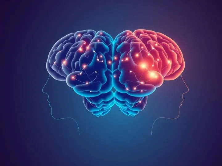
One of the study's key insights is that differences in autism are highly specific, not global. Instead of entire tracts being altered, only certain parts appear to develop differently. These sections often correspond to regions that integrate complex information - such as combining sound and meaning, interpreting facial expressions, or connecting visual input with memory. The researchers note that this pattern may help explain why autistic traits vary widely across individuals: different people may show differences in different portions of the brain's communication pathways.
The study also highlights the value of advanced imaging approaches. The TDF model detected some differences that traditional diffusion tensor imaging did not, suggesting that earlier methods may have missed important details. Similarly, the BUAN approach revealed patterns that would be invisible if measurements were averaged across entire tracts. The combination provides a more precise understanding of how neural pathways develop in autism.
The findings add to a growing view of autism as a condition involving differences in connectivity - how efficiently the brain coordinates information across regions. Prior research has shown variations in brain structure, thickness, and functional activity in autistic individuals, but these patterns often seemed inconsistent or difficult to interpret. This new study suggests that one reason for past inconsistencies may be the limited resolution of older imaging tools. With more refined methods, clearer patterns emerge.
From a practical standpoint, the study does not suggest immediate clinical applications, but it strengthens the scientific foundation for future work. Understanding exactly where structural differences appear may help researchers investigate related behavioral features, such as sensory sensitivity, language development, or social communication differences. It may also guide future studies aiming to understand how these pathways change over time or in response to intervention.
Viewed through Seven Reflections' Dimensional Systems Architecture (DSA) framework, the results illustrate how a complex system can show selective, not uniform, variation. In DSA terms, white matter tracts can be understood as structured pathways that distribute cognitive and sensory information across different "fields" of the brain. Localized differences in specific tract segments indicate that developmental variation affects particular points of high informational load - junctions where different types of processing converge. Rather than representing a global deficit, these findings reveal targeted structural differences within a larger communication system, shaping how information flows and interacts across regions.
By mapping these variations with greater precision, the study moves the field toward a more detailed and nuanced understanding of neurodevelopment in autism. It shows that the brain's wiring architecture can differ in small, specific ways that influence broader patterns of cognition and behavior. As imaging tools continue to advance, studies like this may help bridge the gap between neural structure and real-world experience, offering a clearer picture of how each individual's developmental pathway unfolds.