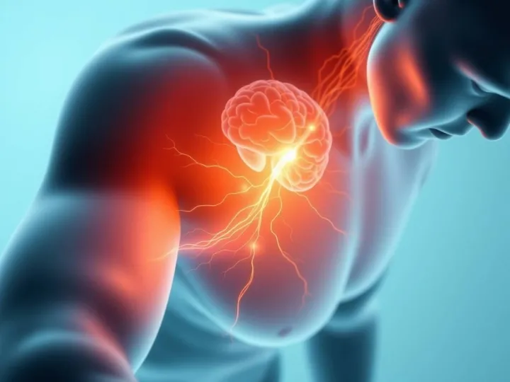
Chronic stress is known to challenge the body's ability to maintain internal balance. When sustained beyond physiological tolerance, it can reconfigure hormonal feedback systems, disrupt neural circuits, and influence vulnerability to addiction. The new research, led by Jalil Rasgado-Toledo and colleagues, examined how long-term stress interacts with alcohol exposure to affect the brain over time. Published in Brain Communications and freely available under a Creative Commons license, the study provides one of the most detailed longitudinal accounts of combined stress-alcohol effects on neurodevelopment.
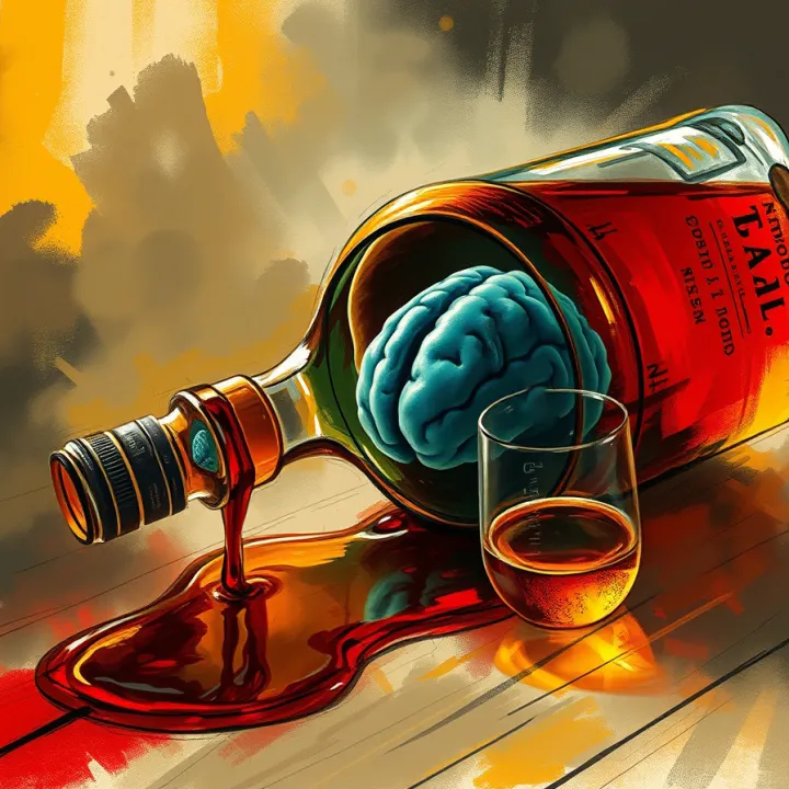
Ninety-six Wistar rats - male and female - were followed from adolescence to adulthood. The animals were assigned to four experimental groups: chronic alcohol exposure, chronic restraint stress, both combined, or control. Stress was applied intermittently through daily three-hour restraint sessions over several weeks, while alcohol was offered via an intermittent access two-bottle-choice protocol providing voluntary 20 percent ethanol intake. Behavioral tests, structural MRI, and resting-state functional MRI were conducted across multiple time points spanning 120 days, roughly the equivalent of a multi-year human developmental window.
Despite expectations, chronic stress did not increase alcohol intake. Males exposed to both stress and alcohol actually showed a slight decline in drinking behavior compared with unstressed drinkers. However, stress alone led to elevated corticosterone levels, slower weight gain, and impaired recognition memory, suggesting systemic strain even in the absence of increased consumption.
Magnetic-resonance imaging revealed more complex consequences. Both chronic stress and alcohol independently produced changes in several key brain regions, and their combined exposure generated additive effects. Reductions in volume were observed in frontal and insular cortices - areas associated with decision-making and interoceptive awareness - along with the hippocampus and amygdala, which regulate memory and emotional response. Conversely, the olfactory bulb, caudate-putamen, entorhinal cortex, and cerebellum displayed volume increases, implying compensatory or maladaptive remodeling.
Sex differences were striking. Male rats exhibited stronger volumetric changes, particularly within subcortical and cerebellar structures, whereas females showed greater alterations in network connectivity. Functional MRI revealed disrupted synchronization across 37 cortical and subcortical regions, including the thalamus, caudate-putamen, hippocampus, and cerebellum. Females displayed intensified coupling between the amygdala and cerebellum as well as the thalamus and motor areas - patterns that may reflect an over-engaged regulatory system under sustained stress.
These findings suggest that stress and alcohol jointly perturb the brain's capacity for coordinated signaling. The structural expansions in some regions may represent swelling or dendritic proliferation linked to overactivity, while shrinkage in others may signal loss of synaptic integrity. Either way, the overall pattern points to a network attempting to rebalance itself through uneven adaptation.
Behavioral outcomes mirrored these physiological changes. Male rats under chronic stress performed poorly in object-recognition tests, indicating memory deficits, while females exposed to alcohol showed slower movement and more cautious exploration - anxiety-like responses without overall changes in standard anxiety indexes. Across groups, locomotor activity correlated with cerebellar and cortical volumes, whereas memory scores correlated with thalamic and olfactory regions.
Crucially, these changes evolved gradually, not as acute reactions. The study's longitudinal design showed that alterations accumulated over weeks, illustrating how repeated low-grade perturbations can reshape developmental trajectories. Instead of catastrophic failure, the system drifted toward a new configuration - one marked by diminished integration and uneven regional compensation.
From the standpoint of neurodevelopment, such slow remodeling can be viewed as entropy accumulation within the brain's organizational field. Each episode of stress or intoxication introduces microscopic disorder - slight mismatches between hormonal timing, metabolic state, and synaptic firing. Over time, these mismatches propagate through interconnected networks, subtly weakening coherence among regions that normally operate in synchrony.
The authors note that this deterioration may underlie the persistent cognitive and emotional difficulties associated with long-term stress or alcohol use disorder. Even when overt behavior appears stable, the underlying network becomes noisier, requiring greater effort to sustain equilibrium. The cerebellum's expansion, for instance, could signify its increased role in compensating for disrupted cortical control - a sign of adaptation that nonetheless burdens the system.
The study also underscores the importance of considering sex as a biological variable. Hormonal fluctuations modulate how stress hormones interact with neurotransmitter systems, producing distinct vulnerabilities. Females showed fewer anatomical losses yet more pronounced functional decoupling - suggesting that structural preservation does not guarantee coherent communication. In contrast, males manifested greater volumetric shifts but less network instability, indicating different modes of adaptation to sustained perturbation.
Viewed through Seven Reflections' Dimensional Systems Architecture (DSA) framework, these findings represent a case of delayed re-integration following sustained entropy input. The brain, conceptualized as a multi-layered field system, maintains coherence by distributing information across structural, functional, and energetic layers. Chronic stress and alcohol act as dual destabilizers: one introduces temporal incoherence via hormonal oscillations, the other adds metabolic and neurochemical noise. Together, they push the system toward a higher-entropy state in which formerly synchronized neural fields begin to drift out of alignment.
In DSA terms, the system's "core coherence field" fragments under prolonged dual load, and the reintegration rate slows. Each attempt at recovery - through synaptic remodeling or neurochemical compensation - creates secondary asymmetries, leading to persistent sub-coherence even after external stressors cease. This delayed re-integration mirrors the behavioral findings: even when drinking behavior normalized, memory and connectivity deficits remained.
Entropy accumulation in neural systems thus manifests not as random chaos but as structured drift - an incremental loss of precise phase relationships among cortical and subcortical oscillators. Restoration of equilibrium requires more than cessation of exposure; it demands re-synchronization across multiple temporal and spatial scales. Future studies could explore how interventions that enhance coherence - whether behavioral, pharmacological, or rhythmic - might accelerate this reintegration process.
By revealing the slow erosion of coherence under chronic stress and alcohol exposure, the Brain Communications study offers a biological illustration of systemic entropy at work. The results suggest that recovery is not merely a matter of detoxification or stress reduction but of re-establishing communication between disjointed neural fields - a process the brain undertakes gradually as it rebuilds its internal order.